Description
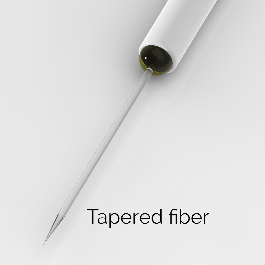
Lambda Fibers are optical fibers that have been tapered down on one end from their full width to a sub-micrometer tip over a length of few millimiters.

Their peculiar optical property is that different optical modes guided by the fiber along its non-tapered portion are out-coupled at different positions along the taper. Depending on how light is launched into the fiber (i.e. how the fiber optical modes are excited), light can be outcoupled from the whole active length of the taper (Full Taper operation) or selectively by different subportions of the active length (Spatial Selective operation).
FULL TAPER OPERATION (Optogenetics and Fiber Photometry)
By exciting all the optical modes of the fiber (i.e. by injecting the light with an optical source having the same or higher Numerical Aperture than the fiber), light will be emitted diffusedly from the taper [1].
This mode of operation is typically employed with Lambda fibers to target large volumes of tissue more homogeneously and efficiently than flat-cut optical fibers [1]. Lambda fibers are typically inserted into the region to be controlled. The tapered profile with sub-micron tip diameter minimizes tissue displacement and insertion damage [1].
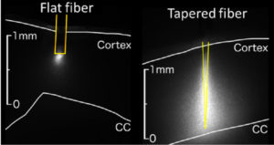
Full taper light emission can be achieved by direct connection of the Lambda fiber to a Laser or LED light source via a patch cable.
Since the optically active surface is larger for a Lambda Fiber than for a flat-cleaved fiber — the surface of a cone with an height equal to the active length vs the fiber core area — a larger total input optical power is needed to obtain the same illumination power density.
The average illumination power density emitted by the active surface of the taper can be calculated as the total light power emitted by the tapered fiber divided by the active area of the tapered fiber. The total light power can be measured by placing the tapered fiber in front and very close to a light power sensor, as is commonly done with flat-cleaved fibers. The active area in squared millimeters can be calculated as the product of the active length of the taper (in millimeters) and a fiber-type dependent coefficient a:
| Fiber type | .22/105 | .39/200 | .66/200 |
| Coefficient a [mm] | 0.086 | 0.189 | 0.243 |
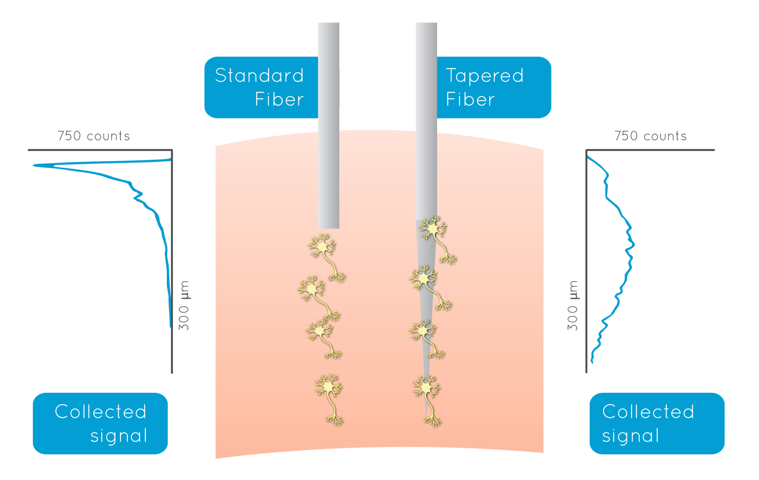
Full taper light collection: Lambda fibers are aso able to collect light all along their active area. By just replacing a standard flat fiber with a Lambda fiber in a conventional fiber photometry experiment, fluorescence is collected nearly homogeneously from large functional regions such as the cortex or the striatum in the mouse. When combined with full taper light delivery, this property translates in a higher signal collected by a Lambda fiber with respect to a flat-cut fiber for similar illumination power density [2].
SPATIAL SELECTIVE OPERATION (Optogenetics and Fiber Photometry)
Light emission can be restricted to sub-portions of the taper by the use of unconventional light delivery methods for exciting only a subset of modes [1]. Emission profiles depend on the Lambda fiber geometry as well as the actual light source employed.
N.B.: This feature can also be employed to correct for implantation errors or uncertainty (Ref. Nature Communications).
This relation between the modal content of the guided light and the active sub-portion of the taper is maintained when using a Lambda fiber for light collection [2], allowing to selectively record light from spatially-confined brain regions at arbitrary depth along the taper. Selectivity is obtained by controlling the position of the excitation, while the elicited fluorescence is collected by the whole taper. This is possible because fluorescence is generated only in the volume of tissue surrounding the taper sub-section selected for delivery of the excitation signal [1].
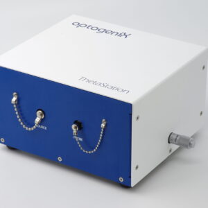
ThetaStation is an entry level, manually operated instrument for spatial selective light delivery with tapered fibers, while GalvoStation is designed for pc controlled spatial selective Optogenetics and Fiber Photometry.
REFERENCES
[1] F. Pisanello, et al., “Dynamic illumination of spatially restricted or large brain volumes via a single tapered optical fiber”, Nature Neuroscience (2017).
[2] F. Pisano, et al., “Depth-resolved fiber photometry with a single tapered optical fiber implant”, Nature Methods (2019)